How Much Radiation Comes From Modern Dental Imaging
How Much Radiation Comes From Modern Dental Imaging?
Modern dental imaging uses much less radiation than many people expect, but the dose still depends on the type of image and why it is being taken. A quick bitewing exam is not the same as a panoramic scan, and neither is the same as cone beam CT.
Key takeaway: Dental X-rays are usually low-dose, and dentists are taught to justify each image rather than take them on a fixed schedule for everyone. According to recent ADA reporting, bitewing X-rays expose a patient to about 5 microsieverts and panoramic X-rays about 20 microsieverts, while cone beam CT can vary much more depending on the machine, field of view, and settings.
Why the answer is “it depends”
People often ask, “How much radiation is a dental X-ray?” as if there is one number. There is not. The American Dental Association overview on X-rays and radiographs makes clear that imaging intervals should be individualized based on age, oral health, risk, and symptoms. More recent ADA coverage of current dental X-ray recommendations notes example doses of roughly 5 microsieverts for bitewings and 20 microsieverts for panoramic imaging, while broader expert recommendations emphasize that cone beam CT spans a much wider range.
A simple dose comparison table
| Imaging type | Typical dose context |
|---|---|
| Bitewing X-rays | about 5 µSv according to recent ADA reporting |
| Panoramic X-ray | about 20 µSv according to recent ADA reporting |
| Cone beam CT | varies widely by field of view and settings; usually higher than routine 2-D dental images |
| Background radiation | everyone receives natural background radiation every day from the environment |
The International Atomic Energy Agency’s dentistry radiation FAQ also places intraoral and cephalometric exams at very low doses, often less than about a day of natural background radiation, while panoramic studies are still relatively low and CBCT may range into tens or hundreds of microsieverts depending on the exam.
Editorial dental imaging room with a panoramic machine and a patient being positioned, emphasizing modern equipment and safety.
What makes one dental image “worth it”
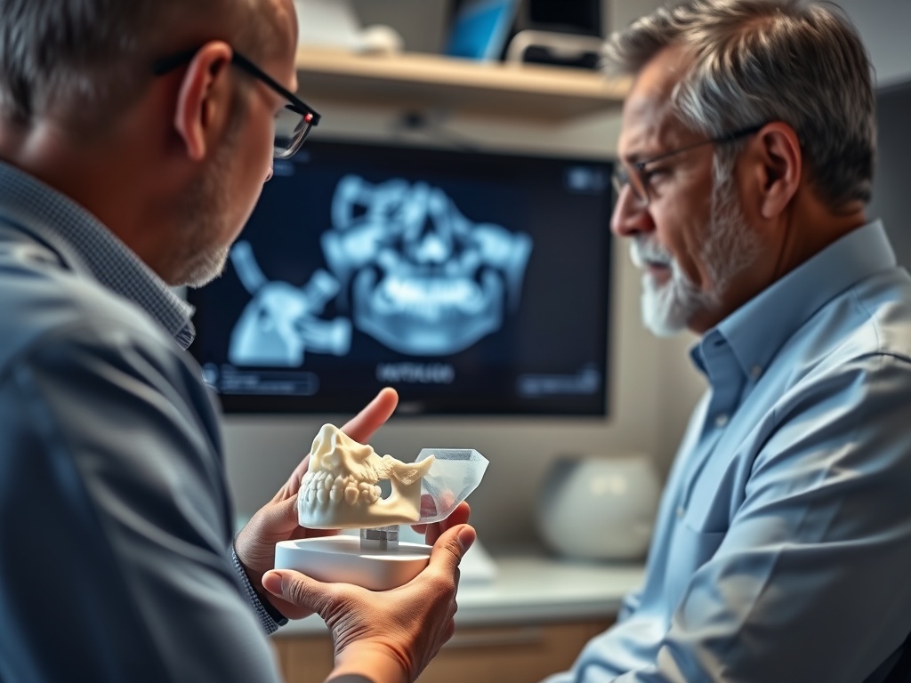
Radiation dose is only half the question. The other half is clinical value. Dentists use imaging when it can reveal disease, bone levels, impacted teeth, infection, root shape, or implant anatomy that cannot be assessed reliably by looking alone. The current expert consensus, reflected in ADA and FDA guidance, is to keep exposure as low as reasonably achievable while still obtaining the image needed for diagnosis.
That means:
- not every visit requires X-rays
- the same person may need different imaging at different life stages
- pregnancy, age, symptoms, and cavity risk all matter
- cone beam CT should be used for specific clinical reasons, not casual curiosity
The questions patients ask most
Are modern dental X-rays safer than older ones?
In general, digital systems and current selection guidelines have reduced exposure compared with older eras, but the main safety point is not “new equals harmless.” It is that imaging should be justified and optimized.
Do I need lead shielding?
Practices follow local regulations and current guidance, but the bigger issue is taking the right image only when needed and using the smallest appropriate field of view, especially for CBCT.
Should I refuse X-rays if I feel fine?
Not automatically. Some dental problems are invisible in a chair exam. A dentist should be able to explain what the image is expected to show and why that information matters for your care.
Planning Notes for Much Radiation Comes Modern Dental Imaging
Routine imaging questions can wait for your next exam if you mainly want to understand your risk and how often images are likely to be recommended. Do not delay care over radiation worries if you have swelling, trauma, severe pain, or signs of infection. In those settings, getting the right image can speed diagnosis and prevent a bigger problem.
If implant treatment is being discussed, imaging is often part of the planning rather than an optional extra. Our article on Implant-Supported Dentures vs Traditional Dentures: Which Feels Better Day to Day? explains why the decision about replacement options often leads to imaging conversations.
If orthodontic planning is the reason images were recommended, our explainer on Why Clear Aligners Need Attachments, Buttons, and Compliance can also help you understand how imaging fits into treatment mechanics.
Questions About Much Radiation Comes Modern Dental Imaging
For Much Radiation Comes Modern Dental Imaging, this part of Questions About Much Radiation Comes Modern Dental Imaging focuses on the symptom history, previous treatment, current medicines, and the result the patient values most after the examination is explained, with the same details verified when the diagnosis is confirmed. The clinician should also explain which examination finding supports each recommendation and what information could change it for Much Radiation Comes Modern Dental Imaging while risks and alternatives are compared, using the examination and health history rather than a general assumption.
Clinical Checks for Much Radiation Comes Modern Dental Imaging
For Much Radiation Comes Modern Dental Imaging, this part of Clinical Checks for Much Radiation Comes Modern Dental Imaging focuses on confirmed findings, measurements, imaging, and health-history factors when a second opinion is considered, with the same details verified when the diagnosis is confirmed. The clinician should also explain what remains uncertain and whether another test or specialist opinion would alter the plan for Much Radiation Comes Modern Dental Imaging when the follow-up plan is written, using the examination and health history rather than a general assumption.
Comparing Options for Much Radiation Comes Modern Dental Imaging
A useful comparison of How Much Radiation Comes From Modern Dental Imaging should also explain what happens if the first choice does not heal, fit, or function as expected. Before choosing a path for How Much Radiation Comes From Modern Dental Imaging, identify which trade-off matters least and which clinical risk matters most before consent is finalized. When comparing approaches to How Much Radiation Comes From Modern Dental Imaging, use the same criteria for every option: expected benefit, limitations, durability, maintenance, timing, and total cost.
A useful way to frame the risk
For most patients, the right question is not “Is there any radiation?” There is. The better question is “Is this image justified, appropriately limited, and likely to change care?” When the answer is yes, the diagnostic benefit often outweighs the low dose of routine dental imaging.
If you are concerned about a recommended image, ask what kind of image it is, what the expected benefit is, and whether a lower-dose or smaller-field option would still answer the clinical question.
Who May Benefit From Much Radiation Comes Modern Dental Imaging
For Much Radiation Comes Modern Dental Imaging, this part of Who May Benefit From Much Radiation Comes Modern Dental Imaging focuses on diagnosis, tissue health, medical history, goals, and ability to maintain the result before temporary care is replaced, with the same details verified when the diagnosis is confirmed. The clinician should also explain which risk factor would make another approach more predictable for Much Radiation Comes Modern Dental Imaging as the treatment sequence is documented, using the examination and health history rather than a general assumption. Recording that explanation gives the patient a clearer next step for Much Radiation Comes Modern Dental Imaging before temporary care is replaced, especially when follow-up duties are reviewed when the diagnosis is confirmed.